


Newsletter
iPSC-Derived Skeletal Muscle; Myoblasts
Applied StemCell (ASC) provides cryopreserved, myoblasts differentiated from an integration-free, control human iPSC line. These high-purity cells express high levels of myoblast biomarkers (CD56, Pax7, Myogenin) and form mature myotubes (skeleton muscle) characterized by elongated, multi-nucleated structures and the expression of myotube marker alpha-MHC in 4-6 days.
- High-purity: ≥90%
- Express Biomarkers: D56, Pax7, Myogenin, and alpha-MHC
- Co-culture Development Service is Available
- GMP iPSC Products & Services >> Learn More
ASC also offers an assortment of fully customizable iPSC differentiation services. With our optimized protocols, we can deliver your fully characterized iPSC-derived cardiomyocytes in just a few weeks. You can send us your iPSCs or we can generate them from you human or non-human samples.
Products and Services
Case Studies
Case Study 1
Characterization of iPSC-derived Myoblasts (ASE-9706)
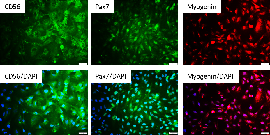
Figure 1. Immunostaining of ASE-9706 iPSC-derived Myoblasts for myoblast biomarkers. Cryopreserved
myoblasts, differentiated from Applied StemCell’s control iPSC line, ASE-9211 were recovered in myoblast culture
media. The cells were stained with myoblast markers, CD56, Pax7, Myogenin on day 2 after recovery. DAPI:
nuclear counterstain; blue. Images at 20x magnification.
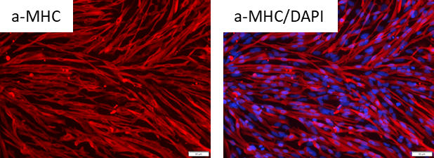
Figure 2. Immunostaining of Myotubes derived from ASE-9706 iPSC-derived Myoblasts for myotube
biomarkers. Cryopreserved myoblasts, differentiated from Applied StemCell’s control iPSC line, ASE-9211 were
recovered in myoblast culture media until they reached confluency and then switched to Myotube Formation
Media for 4 days. The cells were stained with myotube marker, alpha-MHC (a-MHC) on day 4. DAPI: nuclear
counterstain; blue. Images at 20x magnification.