


Newsletter
Cardiomyocytes Differentiation (Ventricular & Atrial)
We provide top-quality service for the directed differentiation of iPSCs into cardiomyocytes using our efficient induction protocol and reagents. We deliver ready-to-use, functional ventricular and atrial cardiomyocytes that are desirable in vitro models for high-content toxicity and drug screening, and cell regeneration research. We deliver receive feasible and physiologically relevant alternatives to embryonic stem cells, primary cells, and animal models.
- High-quality atrial and ventricular cardiomyocytes
- Biomarkers:
- cTNT, alpha-SMA, MLC2A, and MLC2V; video of beating cells
- cTNT, alpha-SMA, MLC2A, and MLC2V; video of beating cells
- We can generate differentiated cardiomyocytes from iPSC derived from healthy or disease patient samples
- Fast turnaround: 2 weeks
- GMP iPSC Differentiation Services Available >> Learn More
Get iPSCs to genetically modify and further differentiate into immune cells? We offer fully customizable iPSC generation from human or non-human tissue samples, and our genome editing experts can use CRISPR or TARGATT™ to genetically alter the iPSCs to fit your project requirements. If you need to expedite your experimental timeline, ASC also offers off-the-shelf cardiomyocytes. Contact us today to find the best solution for your projects.
![]() Beating iPSC-Derived Cardiomyocytes - Video 1| Applied StemCell, Inc.
Beating iPSC-Derived Cardiomyocytes - Video 1| Applied StemCell, Inc.
![]() Beating iPSC-Derived Cardiomyocytes - Video 2 | Applied StemCell, Inc.
Beating iPSC-Derived Cardiomyocytes - Video 2 | Applied StemCell, Inc.
Products and Services
Technical Details

Timelines: 4-6 weeks
- Recovery, Expansion and Validation of iPSCs/ESCs: estimated 3 weeks
- Cardiomyocytes Differentiation: estimated 2-4 weeks
- Characterization of differentiated cells by immunocytochemical (ICC) staining: 2 biomarkers (cTNT; SMA): estimated 2-4 days
Case Studies
Case Study 1:
Characterization Of Ventricular and Atrial Cardiomyocytes
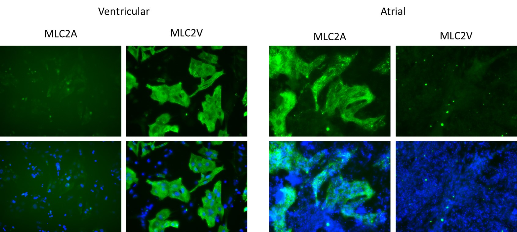
Figure 1. Immunostaining of iPSC-derived ventricular and atrial cardiomyocytes for cardiomyocyte-specific biomarkers.
Cardiomyocytes, differentiated from Applied StemCell’s control iPSC line, ASE-9211 were stained with cardiomyocyte markers, MLC2A and MLC2V.
Support Materials
Brochure: