

Newsletter
Photoreceptor Cell Differentiation
Photoreceptor cells, rods and cones, can be found in the retina and are sensitive to low and high levels of light, respectively. Rods and cones take light and generate signals that are sent to the brain where that information can be processed as part of the first steps in vision. Applied StemCell (ASC) uses non-integrating differentiation protocols to produce photoreceptor cells for your disease modeling, drug target discovery, and other biomedical and applied research. The lineage-committed photoreceptor cells can express photoreceptor-specific markers, including CRX, NR2E3, Tuj1, Rhodopsin, and PPR6a.
To get you the iPSC-derived photoreceptor cells you need, you can send in your healthy or diseased iPSC lines, or you can request to use one of ASC’s control iPSC lines. If you would like to use a genetically engineered iPSC line for the differentiation of photoreceptor cells, ASC also offers high-throughput iPSC genome editing services.
- Optimized differentiation protocols
- We differentiate your healthy or diseased iPSCs or you can select one of ASC's iPSC control lines; iPSC generation is also available
- Standard characterization and additional characterization services are available
- Affordable Price
- Fast turnaround: 3 months
- GMP iPSC Differentiation Services Available >> Learn More
| Photoreceptor Cells | |
| Biomarkers |
Rhodopsin - Standard Service |
| CRX - Inquire | |
| NR2E3 - Inquire | |
| Tuj1 - Inquire | |
| PPR6a - Inquire |
Photoreceptor Differentiation Service Can Include:
- iPSC Generation
- iPSC Characterization
- Photoreceptor Differentiation
- Photoreceptor Characterization
With a fast turnaround time of 12-15 weeks, ASC is the one-stop shop you need to obtain your high-purity photoreceptor cells. We even provide an assortment of assay development services, including Adeno-Associated Virus (AAV) Infectivity assay development (serotype selection, promoter testing, and MOI optimization). Contact us today to speak with one of our experts who can help you design the project you need to drive your research forward.
Products and Services
Case Studies
Case Study 1
iPSC-Derived Photoreceptor Cell Characterization & AAV Transfection
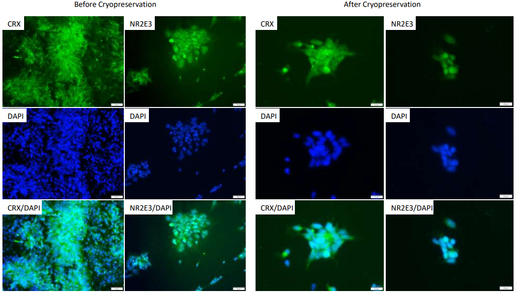
Figure 1: Photoreceptor precursors derived from human iPSCs verified by antibody staining (Left: 20x). The cells with cryopreserved at 1 million cells/vial and recovered in culture media. Antibody staining was completed one day post-thaw (Right: 40x).
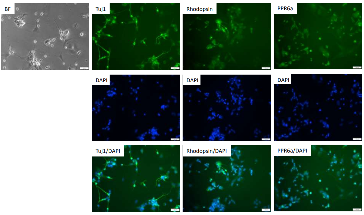
Figure 2: Early-stage iPSC-derived photoreceptor cell antibody staining (two weeks in stage 3 media + 5 days in stage 4 media).
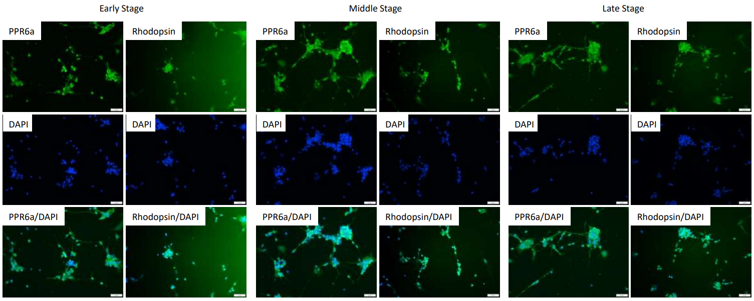
Figure 3: iPSC differentiated photoreceptor cell antibody staining (different time points in stage 3 media + 5 days in stage 4 media). No significant difference was observed at the different time points.
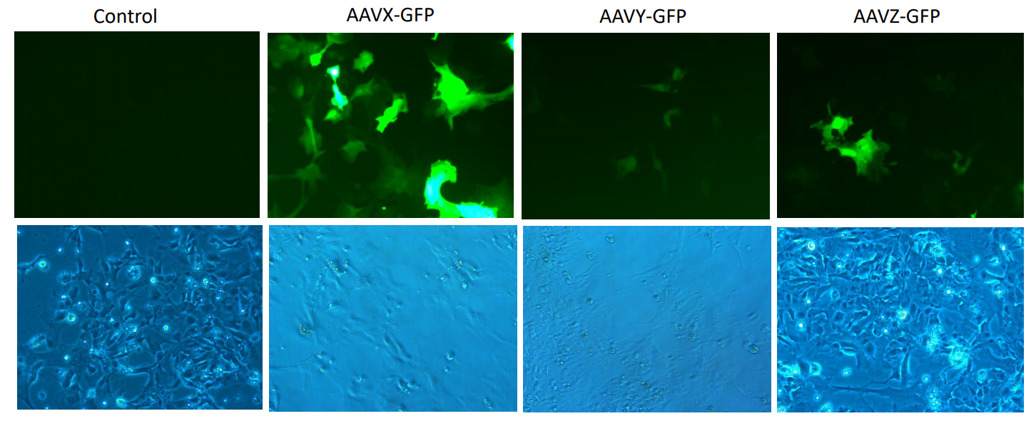
Figure 4: AAV Transfection. 48 hours after virus transduction (seeded 2x10⁵ cells/cell; MOI: 1x10⁵)
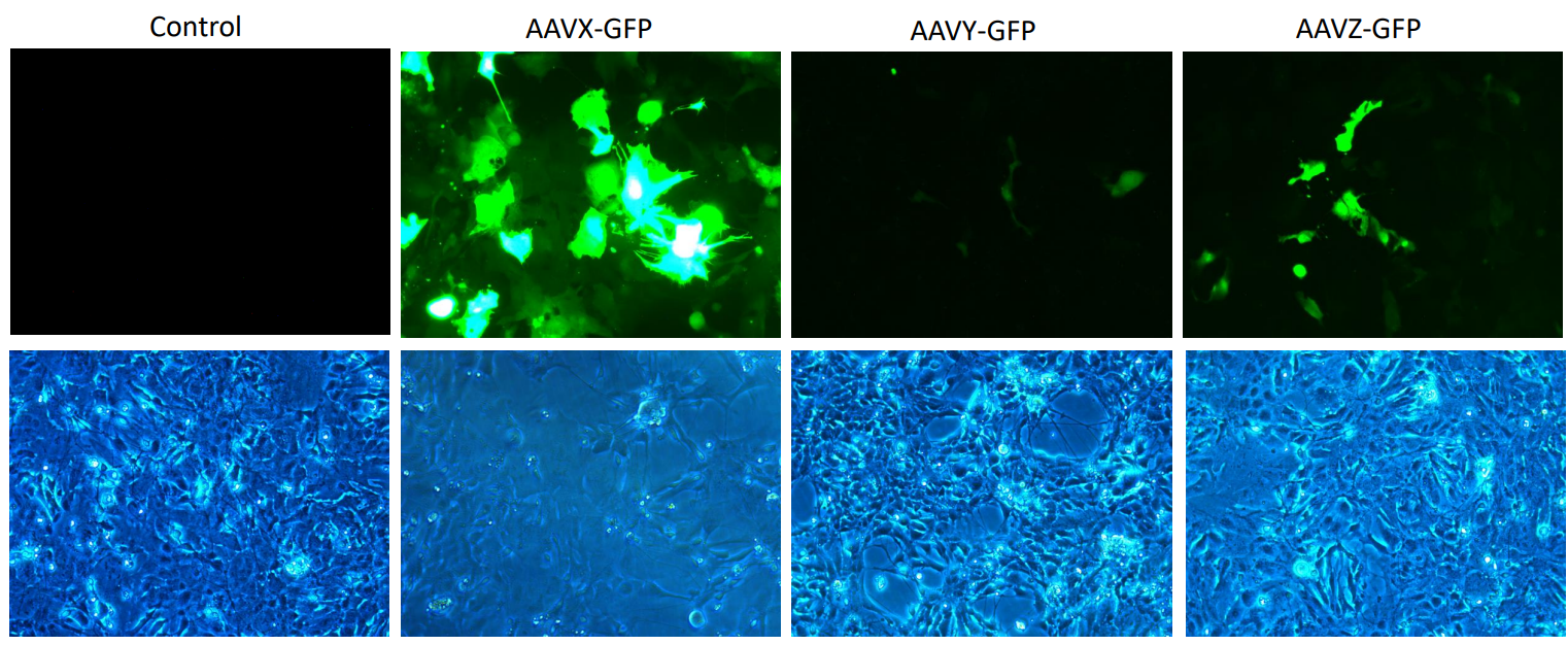
Figure 5: AAV Transfection. 72 hours after virus transduction. (seeded 2x10⁵; MOI: 1x10⁵)