


Newsletter
iPSC-Derived Retinal Pigment Epithelium (RPE) & Photoreceptor Cells
With our optimized differentiation protocols, we produce high-quality cells and deliver well-characterized iPSC-derived photoreceptor and retinal pigment epithelium (RPE) cells for your disease modeling, drug target discovery, and other biomedical and applied research.
iPSC-Derived Photoreceptor Cells
Photoreceptor cells, rods and cones, can be found in the retina and are sensitive to low and high levels of light, respectively. Rods and cones take light and generate signals that are sent to the brain where that information can be processed as part of the first steps in vision. Applied StemCell (ASC) provides off-the-shelf, high-purity iPSC-derived photoreceptor cells that express markers, CRX, NR2E3, Tuj1, Rhodopsin, and PDE6a.
iPSC-Derived Retinal Pigment Epithelium Cells
Retinal Pigment Epithelium (RPE) cells form a single layer in the retina that functions as a selective barrier. RPE can also absorb scattered light and participates in phagocytosis. ASC supplies retinal pigment epithelium (RPE) cells that express markers, PMEL1, MITF, ZO-1, and RPE65.
| Retinal Pigment Epithelium | Photoreceptor Cells | |
| Biomarkers | PMEL1 | CRX |
| MITF | NR2E3 | |
| ZO-1 | Tuj1 | |
| RPE65 | Rhodopsin | |
| PDE6a |
Do you have your own iPSC you would like to differentiate? ASC can use your iPSCs or an ASC Control iPSC Line to produce the photoreceptor or RPE cells you need. We can even develop any assay you may need for your research.
Products and Services
Case Studies
Case Study 1
Characterization of iPSC-derived ASE-9715 Photoreceptor Cells
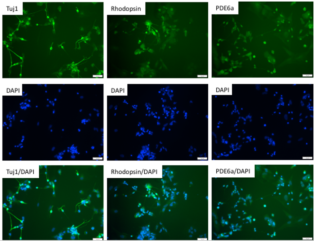
Figure 1. Immunostaining of Mature Photoreceptors derived from human iPSCs for photoreceptor biomarkers. Photoreceptor precursors (ASE-9715), derived from Applied StemCell’s control iPSC line, ASE9211 can be further differentiated in photoreceptor maturation media in 1-2 weeks. The mature photoreceptors were verified by antibody staining with photoreceptor markers, Tuji, Rhodopsin, and PDE6a.
Case Study 2
Characterization of iPSC-derived ASE-9710 RPE Cells
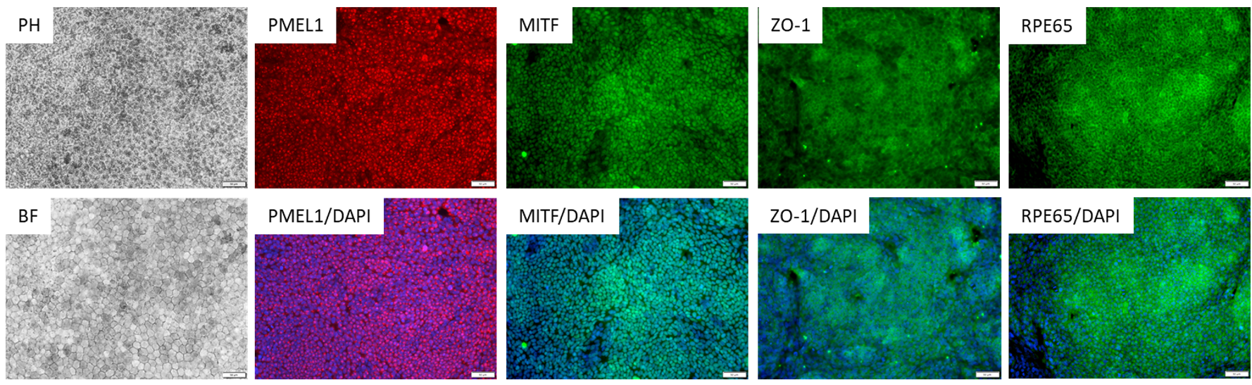
Figure 2. Immunostaining of ASE-9710 iPSC-derived Retinal Pigment Epithelium Cells for retinal pigment
epithelium biomarkers. Cryopreserved RPE cells, differentiated from Applied StemCell’s control iPSC line, ASE9211 were recovered in RPE culture media. The cells were stained with RPE markers, PMEL1, MITF, ZO-1 and
RPE65.
FAQs
What other characterizations have you done on iPSC-RPE (e.g., tight junction measurement, Zo-1 staining, POS phagocytosis, ABCA4 measurement, VEGF and PEDF secretion, retinoid profiling, microvilli at the apical side)?
As specified in the ASE-9710 datasheet, our cells were observed very differently from the picture shown on the datasheet.
Have the scientists done iPSC-RGC cells or other photoreceptor cells? Which type and what characterization?
Photoreceptor Cells: Which percentage are ROD cells? I see a RHO staining but few cells are RHO positive? What cell type are the other cells?
RPE: How many passages can be done in these cells? What is their doubling time? Do you have contamination of other cell types and if yes, in which proportion?
Could we proliferate the photoreceptor cells ?
Are the cells named Photoreceptor Cells (ASE-9715) photoreceptor precursors cells, and media from Applied StemCell (Photoreceptor Maturation Basal Media) differentiate the cell to maturated photoreceptor?
Is there maintenance media for maturated photoreceptors?
Can the maturated photoreceptors be passaged or stocked in cryovials?
RPE: If we decide to try to grow some cells out a passage or two should we just use trypsin to release them or do you recommend some other reagent for detachment?
RPE: What are the recommended cell densities at different well sizes?
In the data sheet it says RPE can be passaged, how many passages are possible?
Maturity of RPE, how long it take to get pigmented cells?
Is there just one media or you recommend different media for RPE maturation?
Any other details you could please share on the RPE?