


Newsletter
iPSC 3D Culture
Applied StemCell (ASC) now provides the tools you need for in vitro 3D induced pluripotent stem cell (iPSC) cultures. The MyEZGel™ iPSC Matrix kit allows the formation of a 3D microenvironment for iPSC growth. With the MyEZGel™ kit, your cells do not have to undergo acidic or chill conditions, and the cells can be easily harvested from the matrix.
ASC also offers a 3D gel of our iPSC control line, ASE-9211. Our ASE-9211 “Master” iPSC line is being used as a benchmark material for developing measurements and standards for the rapidly evolving genetically engineered cells used in cell therapy research. Contact us today to learn more!
Products and Services
Case Studies
Characterization of iPSCs Cultured in MyEZGel™ 3D Matrix
Pluripotency Marker Analysis
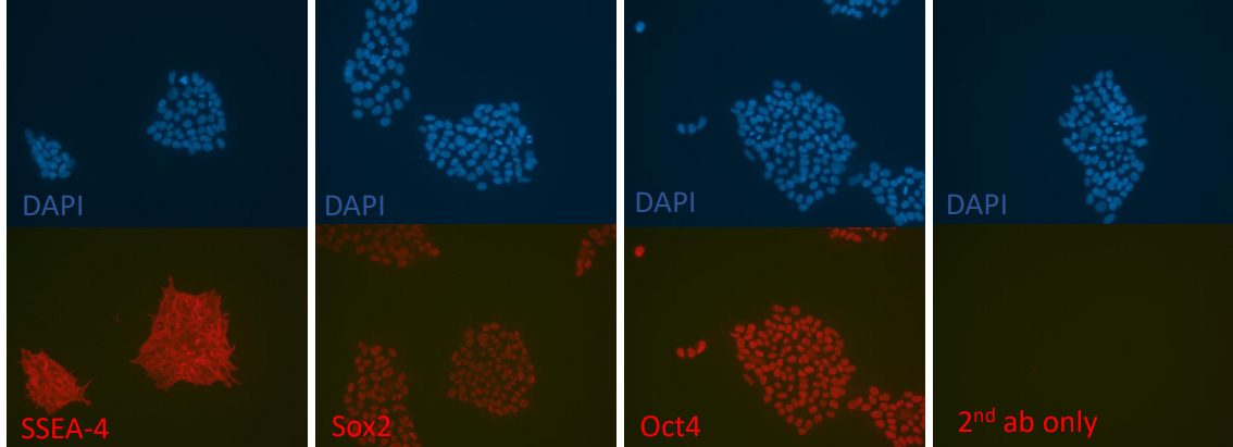
Figure 1. Immunostaining of iPSCs cultured in MyEzGel™ 3D Matrix for pluripotency markers. The 3D-iPSCs expressed common iPSC biomarkers. Image: Nuclear marker: DAPI, Pluripotency markers: SSEA-4, Sox2, Oct4, and 2nd ab only.
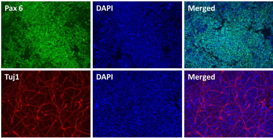
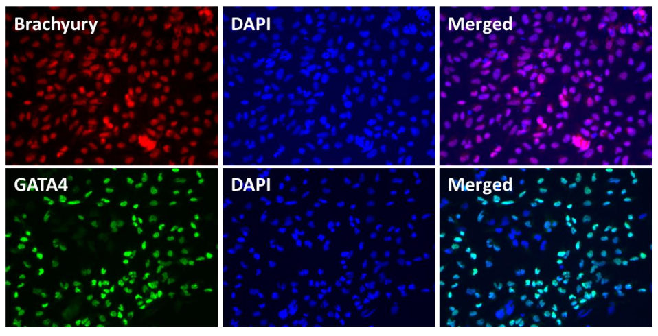
In Vitro Directed Differentiation to the 3 Germ Layers
The iPSCs were recovered and expanded using mTeSR on a Matrigel-coated plate. The cells were disassociated and seeded into a 6-well plate. For the differentiation process, the iPSCs were first treated with Knockout DMEM supplemented with Activin and Wnt3 for Endoderm induction, with BMP4 and Activin for Mesoderm induction, or with Dorsomorphin, SB431542, and Noggin for Ectoderm induction. The staining confirmed the presence of all three germ layers (ectoderm, mesoderm, and endoderm) and the differentiation potential of the 3D cultured iPSCs to all three germ layers.
Ectoderm
Mesoderm
Endoderm
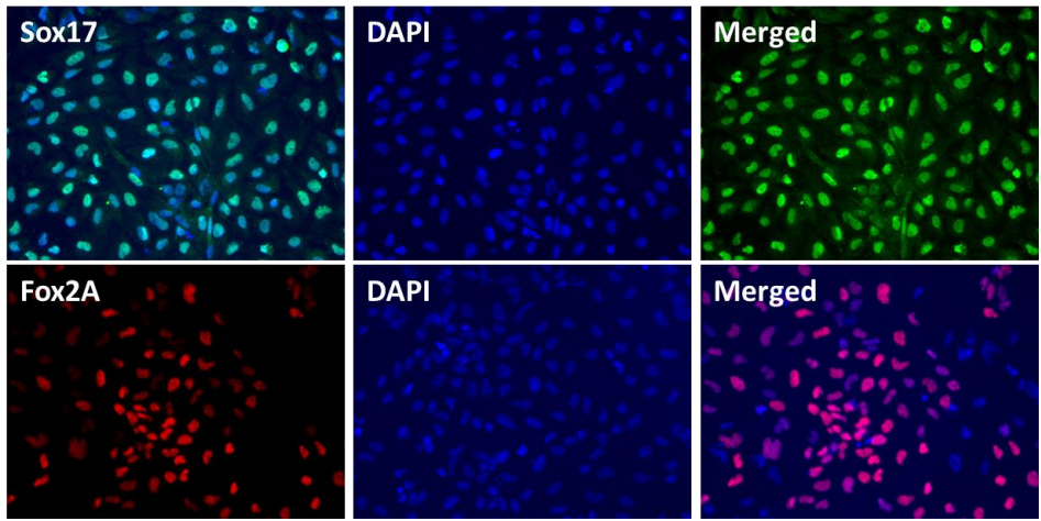
Figure 2. In vitro direct differentiation of MyEZGel™ 3D cultured iPSCs to the three germ layers. Immunofluorescent staining for lineage-specific biomarkers of the 3 germ layers following direct differentiation of the 3D cultured iPSCs. Immunostaining confirmed the differentiation potential of the iPSCs to all 3 germ layers. Images: Nuclear marker: DAPI (blue); Ectoderm markers: Pax6 (green) and Tuji 1 (red); Mesoderm markers: Brachyury (red) and GATA4 (green); Endoderm Sox17 (green-blue) and Fox2A (red).
FAQs
Why do we need to use 0.1% cold water fish gelation in step 4.1.1. ?