Newsletter
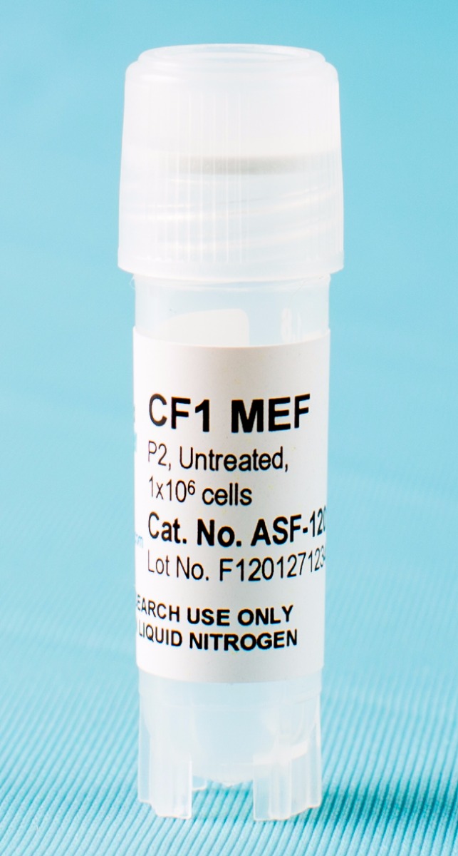
CF-1 MEF Feeder Cells
Derived from CF-1 mouse embryos and used as feeder layers to support the growth of undifferentiated mouse or human embryonic stem cells (ESC) and induced pluripotent stem cells (iPSC) .
MEF cells serve as feeder cells to support the growth of undifferentiated mouse or human embryonic stem cells (ESCs) and induced pluripotent stem cells (iPSCs). MEF cells are isolated from mouse embryos and should be used at an early passage. Before use as feeder cells, MEF cells must be mitotically inactivated by γ-irradiation or mitomycin-C treatment.
CF-1 MEF feeder cells are derived from 13.5 day old mouse embryos and are most commonly used for ESC and iPSC culture.
All MEF cells are manufactured in the US and have an average viability of 95% after thawing. We offer both untreated cells for further expansion and treated cells that can be directly used as a feeder layer.
Also be sure to see our DR4 MEF Feeder Cells, Neo-resistant MEF Feeder Cells, and SNL 76/7 (STO Cell Line) MEF Feeder Cells.

Products and Services
Support Materials
Brochures/ Flyers:
Publications
CF-1 MEF Feeder Cells
- Thakurela, S., Sindhu, C., Yurkovsky, E., Riemenschneider, C., Smith, Z. D., Nachman, I., & Meissner, A. (2019). Differential regulation of OCT4 targets facilitates reacquisition of pluripotency. Nature communications, 10(1), 1-11.
- Smela, M. P., Sybirna, A., Wong, F. C., & Surani, M. A. (2019). Testing the role of SOX15 in human primordial germ cell fate. Wellcome open research, 4.
- Spada, F., Schiffers, S., Kirchner, A., Zhang, Y., Kosmatchev, O., Korytiakova, E., ... & Carell, T. (2019). Oxidative and non-oxidative active turnover of genomic methylcytosine in distinct pluripotent states. BioRxiv, 846584.
- Kiamehr, M., Klettner, A., Richert, E., Koskela, A., Koistinen, A., Skottman, H., ... & Juuti-Uusitalo, K. (2019). Compromised Barrier Function in Human Induced Pluripotent Stem-Cell-Derived Retinal Pigment Epithelial Cells from Type 2 Diabetic Patients. International journal of molecular sciences, 20(15), 3773.
- Barber, K., Studer, L., & Fattahi, F. (2019). Derivation of enteric neuron lineages from human pluripotent stem cells. Nature protocols, 14:1261–1279.
- Berecz, T., Husvéth-Tóth, M., Mioulane, M., Merkely, B., Apáti, Á., & Földes, G. (2019). Generation and Analysis of Pluripotent Stem Cell-Derived Cardiomyocytes and Endothelial Cells for High Content Screening Purposes. In: Methods in Molecular Biology. Humana Press.
- Madak-Erdogan, Z., Band, S., Zhao, Y. C., Smith, B. P., Kulkoyluoglu-Cotul, E., Zuo, Q., ... & Kim, S. H. (2019). Free fatty acids rewire cancer metabolism in obesity-associated breast cancer via estrogen receptor and mTOR signaling. Cancer research, canres-2849.
- Deuse, T., Hu, X., Gravina, A., Wang, D., Tediashvili, G., De, C., ... & Davis, M. M. (2019). Hypoimmunogenic derivatives of induced pluripotent stem cells evade immune rejection in fully immunocompetent allogeneic recipients. Nature biotechnology, 1.
- Kiamehr, M. (2019). Induced pluripotent stem cell-derived hepatocyte-like cells: The lipid status in differentiation, functionality, and de-differentiation of hepatic cells. Tampere University Dissertations.
- Yeom, K. H., Mitchell, S., Linares, A. J., Zheng, S., Lin, C. H., Wang, X. J., ... & Black, D. L. (2018). Polypyrimidine Tract Binding Protein blocks microRNA-124 biogenesis to enforce its neuronal specific expression. bioRxiv, 297515. https://doi.org/10.1101/297515
- Chai, S., Wan, X., Ramirez-Navarro, A., Tesar, P. J., Kaufman, E. S., Ficker, E., ... & Deschênes, I. (2018). Physiological genomics identifies genetic modifiers of long QT syndrome type 2 severity. The Journal of Clinical Investigation, 128(3). DOI: 10.1172/JCI94996
- Oh, Y., Zhang, F., Wang, Y., Lee, E. M., Choi, I. Y., Lim, H., ... & Wu, H. (2017). Zika virus directly infects peripheral neurons and induces cell death. Nature Neuroscience, 20(9), 1209-1212.
- Kiamehr, M., Viiri, L. E., Vihervaara, T., Koistinen, K. M., Hilvo, M., Ekroos, K., ... & Aalto-Setälä, K. (2017). Lipidomic profiling of patient-specific induced pluripotent stem cell-derived hepatocyte-like cells. Disease Models & Mechanisms, dmm-030841.
- Wong, K. G., et al. (2017). CryoPause: A New Method to Immediately Initiate Experiments after Cryopreservation of Pluripotent Stem Cells. http://www.cell.com/stem-cell-reports/pdfExtended/S2213-6711(17)30217-5.
- Cvetkovic, C., et al. (2017). A 3D-printed platform for modular neuromuscular motor units. Microsystems & Nanoengineering, 3, 17015.
- Kurapati, S., et al. (2017). Role of JNK pathway in varicella-zoster virus lytic infection and reactivation. Journal of Virology, JVI-00640.
- Kotini, A. G., Chang, C. J., Chow, A., Yuan, H., Ho, T. C., Wang, T., ... & Teruya-Feldstein, J. (2017). Stage-specific human induced pluripotent stem cells map the progression of myeloid transformation to transplantable leukemia. Cell Stem Cell, 20(3), 315-328.
- Maghen, L., Shlush, E., Gat, I., Filice, M., Barretto, T. A., Jarvi, K., ... & Librach, C. L. (2017). Human umbilical perivascular cells (HUCPVCs): a novel source of mesenchymal stromal-like (MSC) cells to support the regeneration of the testicular niche. Reproduction, 153(1), 85-95.
For more references, visit our reference page.
FAQs
What is the maximum number of passages for healthy primary MEFs culture? (ASF-1201)


-L1_L2.jpg)
